Back Bones Diagram - Diagram Of Vertebral Column Showing Different Parts And Regions Of The Download Scientific Diagram - The vast difference in height and limb length between birth and adulthood are mainly the result of endochondral ossification in the.
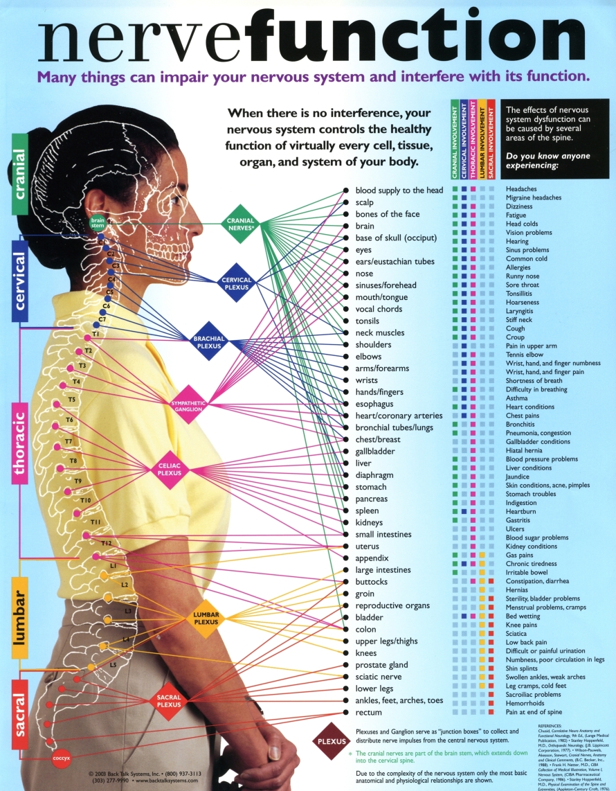
Back Bones Diagram - Diagram Of Vertebral Column Showing Different Parts And Regions Of The Download Scientific Diagram - The vast difference in height and limb length between birth and adulthood are mainly the result of endochondral ossification in the.. The first seven bones (vertebrae) of your spine form your neck. It is designed to be incredibly strong, protecting the highly sensitive nerve roots, yet highly flexible, providing for mobility on many different planes. Bone structure wide hips 12 photos of the bone structure wide hips bone structure wide hips, bone, bone structure wide hips These problems can reduce the amount of space available for your spinal cord and the nerves that branch off it. The lower back region contains large muscles that support the back and allow for movement in the trunk of the body.
The sciatic nerve is the dominant nerve that innervates the lower back and the lower extremities. This article looks at the anatomy of the back, including bones, muscles, and nerves. Other important bones in the shoulder include: Individual anatomical structures include 2: This bone is on the thumb side of the hand near the radius.;

Bones, discs, and joints in your lower back.
Sciatica occurs when the sciatic nerve becomes pinched, usually by a herniated disk in your spine or by an overgrowth of bone (bone spur) on your vertebrae. Bones in the body 12 photos of the bones in the body bones in the body and what they do, bones in the body chart, bones of the body book, bones of the body made easy, remembering bones in the body, bone, bones in the body and what they do, bones in the body chart, … The cranial bones include occipital bone, two parietal bones, frontal bone, two temporal bones, sphenoid bone, and the ethmoid bone. At the back of each bone in the spine (vertebra) are bony points called processes, which muscles attach to. The lower bone density test results, the lower your total bone density, indicating osteopenia or osteoporosis. The human skeleton, like that of other vertebrates, consists of two principal subdivisions, each with origins distinct from the others and each presenting certain individual features.these are (1) the axial, comprising the vertebral column—the spine—and much of the skull, and (2) the appendicular, to which the pelvic (hip) and pectoral (shoulder) girdles and the bones and cartilages of the. Many of the nerves of the peripheral nervous system, or pns, branch out from the spinal cord and travel to various. A tough, springy disc of cartilage sits between the vertebrae of your spine. This bone is on the thumb side of the hand near the radius.; It travels from the lower spine, through the pelvis, and down each leg. Human back bones diagram poster 28 inch x 24 inch 16 inch x 13 inch. Spinal anatomy is a remarkable combination of strong bones, flexible ligaments and tendons, large muscles and highly sensitive nerves. The first seven bones (vertebrae) of your spine form your neck.
The cranial bones include occipital bone, two parietal bones, frontal bone, two temporal bones, sphenoid bone, and the ethmoid bone. The spine diagram the spine diagram shown below, consists of many bones or vertebrae,soft discs,the spinal cord, and spinal nerves. A tough, springy disc of cartilage sits between the vertebrae of your spine. This article looks at the anatomy of the back, including bones, muscles, and nerves. Long bone diagram labeled colored.
The red lines point individual bones and the names are writen in singular, the blue lines conect to group of bones and are in plural form.
The lower bone density test results, the lower your total bone density, indicating osteopenia or osteoporosis. The spinal cord begins at the base of the brain and extends into the pelvis. Anatomical diagrams of the spine and back. The eight bones of the wrist are:. These muscles can spasm or become strained, which is a common cause of lower back pain. The fourth lumbar spine vertebra (l4) is located towards the bottom of the lumbar section, near the sacral vertebrae at the bottom of the spine.like the other lumbar spinal bones, l4 primarily. One way to learn all the bones in the human body is to categorize them by shape. Lateral labeled diagram of the human vertebral spinal column showing vertebrae numbering order and the 5 different regions of the spine. Spinal anatomy is a remarkable combination of strong bones, flexible ligaments and tendons, large muscles and highly sensitive nerves. While in the thoracic and lumbar spine, the anatomy of the vertebrae is relatively consistent between each vertebra, cervical spine anatomy is quite variable. The atlas is the topmost vertebra, and along with c2, forms the joint connecting the skull and spine. Bones in the body 12 photos of the bones in the body bones in the body and what they do, bones in the body chart, bones of the body book, bones of the body made easy, remembering bones in the body, bone, bones in the body and what they do, bones in the body chart, … Other important bones in the shoulder include:
Bones prevent you from puddling on the floor in the form of a jellyfish, but what else do they do?. Diagram of a human female skeleton, back view. The fourth lumbar spine vertebra (l4) is located towards the bottom of the lumbar section, near the sacral vertebrae at the bottom of the spine.like the other lumbar spinal bones, l4 primarily. These muscles can spasm or become strained, which is a common cause of lower back pain. The first seven bones (vertebrae) of your spine form your neck.

The cervical spine has 7 stacked bones called vertebrae, labeled c1 through c7.
Bones prevent you from puddling on the floor in the form of a jellyfish, but what else do they do?. The shoulder joint is formed where the humerus (upper arm bone) fits into the scapula (shoulder blade), like a ball and socket. This bone is on the thumb side of the hand near the radius.; These bones work together to provide. Bones, discs, and joints in your lower back. The fourth lumbar spine vertebra (l4) is located towards the bottom of the lumbar section, near the sacral vertebrae at the bottom of the spine.like the other lumbar spinal bones, l4 primarily. One way to learn all the bones in the human body is to categorize them by shape. The red lines point individual bones and the names are writen in singular, the blue lines conect to group of bones and are in plural form. Related posts of anatomy of the back organs women abdominal anatomy. As viewed from the side, the cervical spine forms a lordotic curve by gently curving toward the front of the body and then back. The top of the cervical spine connects to the skull, and the bottom connects to the upper back at about shoulder level. This article looks at the anatomy of the back, including bones, muscles, and nerves. Bone structure wide hips 12 photos of the bone structure wide hips bone structure wide hips, bone, bone structure wide hips
Komentar
Posting Komentar